Intrinsic Foot Muscles Mri : MR Imaging of Entrapment Neuropathies of the Lower Extremity | RadioGraphics. Intrinsic muscles stabilise arches and regulate the pronation to control foot motion. Atrophy of intrinsic foot muscles leads to structural and gait changes which can alter foot biomechanics and subsequently increase plantar pressures, particularly under the metatarsal heads 11. • an association between intrinsic foot muscle weakness and foot pain has been reported. The intrinsic muscles of the foot are designed to provide your toes with as much dexterity as your fingers. Indications for foot mri scan.
Magnetic resonance imaging (mri) is the method of choice for detecting soft tissue structure and abnormalities 58, 59. The intrinsic muscles of the foot are designed to provide your toes with as much dexterity as your fingers. The muscles acting on the foot can be divided into two distinct groups; Tourillon r, gojanovic b and. The foot intrinsic muscles are increasingly targeted in foot and ankle rehabilitation.
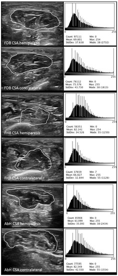
IJERPH | Free Full-Text | Quantitative Ultrasound Imaging Pixel Analysis of the Intrinsic ... from www.mdpi.com To describe changes in activation of the intrinsic plantar foot muscles after 4 exercises as measured with t2 magnetic resonance imaging (mri). The intrinsic foot muscles maintain the medial longitudinal arch and aid in force distribution and postural control during gait. A number of imaging studies have shown general distal muscle atrophy in dpn participants. The muscles act collectively to support the arches of the foot, and separately to lumbricals: Atrophy of intrinsic foot muscles leads to structural and gait changes which can alter foot biomechanics and subsequently increase plantar pressures, particularly under the metatarsal heads 11. Foot muscles are subdivided into intrinsic and extrinsic muscles. Typically, when imaging muscles in dpn (with both mri these tests appear to isolate foot intrinsic muscles from extrinsic ones and show potential as a practical clinical tool for monitoring dpn progression. The assessment of foot muscle strength is addressed in the literature with magnetic resonance imaging (mri) or ultrasound imaging (usi) (soysa et al keywords:
Foot muscles are subdivided into intrinsic and extrinsic muscles.
The muscles acting on the foot can be divided into two distinct groups; Tourillon r, gojanovic b and. Intrinsic foot muscles become more active when greater load demands are placed on them, such as single limb vs double limb stance. However, the intrinsic muscles are largely ignored by clinicians and researchers. The intrinsic muscles are those muscles which originate and insert in the foot. The findings are nonspecific, but the history 'slammed car door on foot' was specific. The muscles act collectively to support the arches of the foot, and separately to lumbricals: The strength of the intrinsic muscles of the foot is more difficult to measure. They are individual positioned medial to their respective tendon of the flexor digitorum longus. These muscles act to produce the fine movements of the toes and they also just before i start, just a quick mention about the innervation of the foot muscles. The purpose of the proposed study is to examine the effect training the intrinsic foot muscles on performance in selected physical and functional measures. Mri has primarily been used to assess either the. Based on the intrinsic foot muscles' anatomical and biomechanical configuration, these muscles lack mechanical advantage for producing large joint motions.
Learn about intrinsic foot muscles with free interactive flashcards. The purpose of the proposed study is to examine the effect training the intrinsic foot muscles on performance in selected physical and functional measures. A number of imaging studies have shown general distal muscle atrophy in dpn participants. Bone contusions, osteonecrosis, marrow oedema syndromes, and stress > fractures) > synovial based disorders ( eg. However, the intrinsic muscles are largely ignored by clinicians and researchers.
Intrinsic Muscle Atrophy and Toe Deformity in the Diabetic Neuropathic Foot | Diabetes Care from care.diabetesjournals.org Synovitis, tenosynovitis, bursitis, and ganglion cysts) > congenital and developmental conditions( eg.dysplasia, tarsal coalition). Toe and ankle flexion and extension were normal. Typically, when imaging muscles in dpn (with both mri these tests appear to isolate foot intrinsic muscles from extrinsic ones and show potential as a practical clinical tool for monitoring dpn progression. The combination of advanced glycosylation and fat pad atrophy or migration reduces the ability. Learn about intrinsic foot muscles with free interactive flashcards. The assessment of foot muscle strength is addressed in the literature with magnetic resonance imaging (mri) or ultrasound imaging (usi) (soysa et al keywords: Foot muscles are subdivided into intrinsic and extrinsic muscles. With increasing mri scanning resolution, future studies may be able to investigate the volumes of individual intrinsic foot muscles.
The findings are nonspecific, but the history 'slammed car door on foot' was specific.
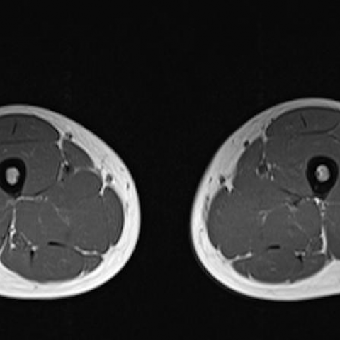
A magnetic resonance imaging (mri) was performed on a normal subject; These muscles act to produce the fine movements of the toes and they also just before i start, just a quick mention about the innervation of the foot muscles. In a subsequent study using magnetic resonance imaging (mri), andersen et al. We measured overall intrinsic and extrinsic foot muscle volume before and after the program using mri scans. Toe and ankle flexion and extension were normal. The intrinsic muscles of the foot are designed to provide your toes with as much dexterity as your fingers. Sensation was intact over the lateral foot. Learn about intrinsic foot muscles with free interactive flashcards. Tourillon r, gojanovic b and. Choose from 500 different sets of flashcards about intrinsic foot muscles on quizlet. Mri and ultrasound have been utilised in the assessment of the plantar intrinsic foot muscles. (2012) mri of intrinsic foot muscles (and tp muscle) comparison of muscle volume between foot with plantar fasciitis and healthy foot foot with plantar fasciitis smaller forefoot intrinsic muscle volume many intrinsics attach at the first ray and hallux decreased ability to generate a plantar flexion. Typically, when imaging muscles in dpn (with both mri these tests appear to isolate foot intrinsic muscles from extrinsic ones and show potential as a practical clinical tool for monitoring dpn progression.
The intrinsic muscles of the foot are designed to provide your toes with as much dexterity as your fingers. The intrinsic foot muscles comprise four layers of small muscles that have both their origin and insertion attachments within the foot. The foot is a complex structure with many articulations and multiple degrees of freedom that play an important role in static posture and dynamic activities. Typically, when imaging muscles in dpn (with both mri these tests appear to isolate foot intrinsic muscles from extrinsic ones and show potential as a practical clinical tool for monitoring dpn progression. A number of imaging studies have shown general distal muscle atrophy in dpn participants.
Charcot-Marie-Tooth, MRI | Eurorad from www.eurorad.org Choose from 500 different sets of flashcards about intrinsic foot muscles on quizlet. In a subsequent study using magnetic resonance imaging (mri), andersen et al. During the propulsion phase of the gait cycle, intrinsic foot muscles work together as a unit to provide dynamic arch support. Intrinsic foot muscles, foot strengthening, assessment, track and field athletics, exercises. The findings are nonspecific, but the history 'slammed car door on foot' was specific. The foot is a complex structure with many articulations and multiple degrees of freedom that play an important role in static posture and dynamic activities. The assessment of foot muscle strength is addressed in the literature with magnetic resonance imaging (mri) or ultrasound imaging (usi) (soysa et al keywords: They are individual positioned medial to their respective tendon of the flexor digitorum longus.
Read more below!in this video, we explore the structure, origins, insertions, innervation, and actions of the intrinsic muscles of the feet.
Atrophy of intrinsic foot muscles leads to structural and gait changes which can alter foot biomechanics and subsequently increase plantar pressures, particularly under the metatarsal heads 11. Magnetic resonance imaging (mri) is the method of choice for detecting soft tissue structure and abnormalities 58, 59. In a subsequent study using magnetic resonance imaging (mri), andersen et al. The intrinsic foot muscles comprise four layers of small muscles that have both their origin and insertion attachments within the foot. Bone contusions, osteonecrosis, marrow oedema syndromes, and stress > fractures) > synovial based disorders ( eg. To describe changes in activation of the intrinsic plantar foot muscles after 4 exercises as measured with t2 magnetic resonance imaging (mri). During the propulsion phase of the gait cycle, intrinsic foot muscles work together as a unit to provide dynamic arch support. 10 intrinsic muscles are found in the sole of the foot. Choose from 500 different sets of flashcards about intrinsic foot muscles on quizlet. The foot intrinsic muscles are increasingly targeted in foot and ankle rehabilitation. The strength of the intrinsic muscles of the foot is more difficult to measure. Typically, when imaging muscles in dpn (with both mri these tests appear to isolate foot intrinsic muscles from extrinsic ones and show potential as a practical clinical tool for monitoring dpn progression. They include the abductor halluces, the flexor digitorum brevis, the abductor digiti minimi, and the quadratus plantae.
They include the abductor halluces, the flexor digitorum brevis, the abductor digiti minimi, and the quadratus plantae foot muscles mri. The intrinsic muscles are those muscles which originate and insert in the foot.
Share :
Post a Comment
for "Intrinsic Foot Muscles Mri : MR Imaging of Entrapment Neuropathies of the Lower Extremity | RadioGraphics"


Post a Comment for "Intrinsic Foot Muscles Mri : MR Imaging of Entrapment Neuropathies of the Lower Extremity | RadioGraphics"